
In the accelerating global energy transition, the quest for efficient, low-cost, and easily manufacturable photovoltaic technologies has never been more critical. Among the most promising candidates are next-generation thin film solar panels, particularly those based on organic and perovskite semiconductors. These technologies have captivated the research community due to their potential for solution-processed fabrication, compatibility with flexible substrates, and their remarkably rapid efficiency improvements. Laboratory-scale organic photovoltaic (OPV) cells have now surpassed 20% power conversion efficiency (PCE), while perovskite solar cells (PSCs) have exceeded 26%. However, the path from high-efficiency small-area devices to reliable, large-area panels is fraught with challenges, primarily concerning uniformity and long-term stability. The performance of these thin film solar panels is exquisitely sensitive to micro-scale defects and inhomogeneities that arise during fabrication and operational degradation. Therefore, advanced characterization techniques capable of mapping these defects are indispensable for both fundamental understanding and technological advancement.
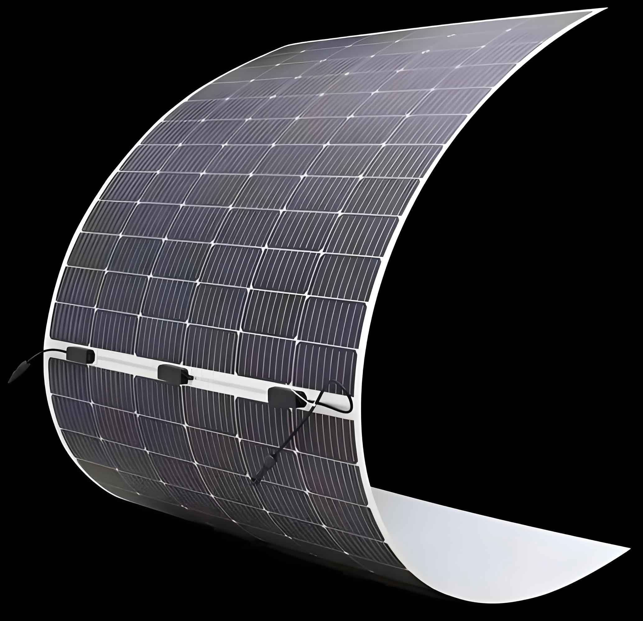
The core of a thin film solar panel is a multilayer “sandwich” structure. Light enters through a transparent conductive oxide (TCO) bottom electrode, such as indium tin oxide (ITO). The heart of the device is the photoactive layer, where photons are absorbed to create excitons (bound electron-hole pairs) which subsequently dissociate into free charge carriers. In OPVs, this layer typically consists of a bulk heterojunction (BHJ) blend of electron-donor and electron-acceptor organic semiconductors. In PSCs, it is a film of ABX3 perovskite semiconductor (e.g., CH3NH3PbI3). On either side of the active layer, charge transport layers (CTLs) facilitate the selective extraction of holes and electrons to the respective electrodes—a metal top electrode (e.g., Ag, Al) and the TCO bottom electrode. The efficiency of this entire process can be encapsulated by the power conversion efficiency formula:
$$PCE = \frac{J_{sc} \times V_{oc} \times FF}{P_{in}}$$
where $J_{sc}$ is the short-circuit current density, $V_{oc}$ is the open-circuit voltage, $FF$ is the fill factor, and $P_{in}$ is the incident light power. Any defect within or between these nanoscale layers directly degrades one or more of these parameters, undermining the performance of the thin film solar panel.
Defects in these panels are multifaceted. They can be intrinsic, such as pinholes in the active layer, poor crystallinity, or compositional inhomogeneity in perovskite films. They can also be extrinsic, like dust particles incorporated during processing, local delamination of layers, or corrosion of electrodes. Interfacial defects, where charge extraction occurs, are particularly detrimental as they promote non-radiative recombination, quantified by the relation for open-circuit voltage loss:
$$\Delta V_{oc} = \frac{n k_B T}{q} \ln\left(1 + \frac{J_{bi}}{J_{0}}\right)$$
where $n$ is the ideality factor, $k_B$ is Boltzmann’s constant, $T$ is temperature, $q$ is the elementary charge, $J_{bi}$ is the bimolecular recombination current, and $J_{0}$ is the reverse saturation current, which is heavily influenced by defect density. Locating, identifying, and understanding these defects require spatially resolved imaging techniques that go beyond standard device-averaging measurements.
Microscale Defect Imaging: Principles and Methodologies
To probe the health of a thin film solar panel at the micron scale, several non-destructive imaging techniques have been adapted and developed. Each method probes a different physical mechanism, providing complementary information about the nature and location of defects.
Photoluminescence (PL) Imaging
PL imaging involves exciting the photoactive layer with a specific wavelength of light and mapping the resulting emission intensity and/or lifetime. In a semiconductor, photoexcitation promotes electrons to the conduction band, leaving holes in the valence band. These photogenerated carriers can recombine radiatively, emitting a photon. The probability of radiative recombination is strongly suppressed by the presence of defect states that provide non-radiative pathways. Therefore, areas with high defect density appear dark in a PL intensity map. The local implied open-circuit voltage ($iV_{oc}$) can be extracted from the PL intensity ($I_{PL}$) using the generalized Planck’s law relation:
$$I_{PL} \propto \exp\left(\frac{q \cdot iV_{oc}}{k_B T}\right)$$
This makes PL imaging a powerful tool for assessing the electronic quality and homogeneity of perovskite and organic semiconductor films, especially before complete device fabrication. It is highly sensitive to bulk and interfacial non-radiative recombination centers within the light-absorbing layer itself.
Electroluminescence (EL) Imaging
EL imaging is the inverse process: an external voltage or current is applied to the solar cell in the dark, injecting electrons and holes that then recombine radiatively within the active layer. The emitted light is captured to form an image. The EL signal intensity $I_{EL}$ is related to the injected current density $J$ and the internal radiative efficiency $\eta_{int}$:
$$I_{EL} \propto \eta_{int} \cdot J \cdot \exp\left(\frac{qV}{nk_B T}\right)$$
Areas with high series resistance (e.g., poor electrode contact), shunts, or regions where charge injection is blocked will show reduced or no EL emission. Thus, EL imaging is exceptionally sensitive to electrically active defects, particularly those related to charge injection and extraction at the interfaces and contacts. It directly visualizes the functionality of the complete device stack under operating (injection) conditions.
Laser Beam Induced Current (LBIC) Imaging
LBIC is arguably the most direct functional mapping technique for a thin film solar panel. A focused laser beam (spot size can be down to ~1 µm) is raster-scanned across the device. At each point, the generated photocurrent is measured while the device is held at short-circuit or a defined bias. The measured current $I_{LBIC}$ at position $(x,y)$ is:
$$I_{LBIC}(x,y) = q \cdot \Phi \cdot EQE(x,y) \cdot G(x,y)$$
where $q$ is the elementary charge, $\Phi$ is the photon flux of the laser, $EQE(x,y)$ is the local external quantum efficiency, and $G(x,y)$ is a factor accounting for charge collection efficiency. LBIC directly maps the spatial variation in the device’s ability to convert light into extractable current. It is sensitive to all types of defects that affect light absorption, charge generation, or charge collection, including shunts, series resistance variations, and active layer inhomogeneities.
Lock-in Thermography (LIT)
LIT detects local heating effects caused by defects. A periodic electrical stimulus (current or voltage) is applied to the device, and an infrared camera synchronized to this “lock-in” frequency maps the resulting temperature variations. In dark LIT (DLIT), the heat generated by unwanted shunt currents or areas of high series resistance is detected. In illuminated LIT (ILIT), the cell is under light, and the technique can map the distribution of recombination heat. The local temperature rise $\Delta T$ is proportional to the local power dissipation $P_{diss}$:
$$\Delta T(x,y) \propto P_{diss}(x,y) = I_{sh}(x,y)^2 \cdot R_{sh}(x,y)$$
where $I_{sh}$ is the shunt current and $R_{sh}$ is the shunt resistance. LIT is unparalleled in pinpointing the exact location of strong shunts and hot spots that can lead to catastrophic failure in a thin film solar panel module.
The characteristics of these primary imaging techniques are summarized in the table below.
| Imaging Technique | Physical Principle | Primary Information | Spatial Resolution | Key Measurable |
|---|---|---|---|---|
| Photoluminescence (PL) | Light excitation → Radiative recombination | Material quality, defect density, $iV_{oc}$ map | < 1 µm | PL Intensity, Lifetime |
| Electroluminescence (EL) | Charge injection → Radiative recombination | Electrical integrity, contact issues, shunt/resistance maps | ~10-50 µm | EL Intensity |
| Laser Beam Induced Current (LBIC) | Focused light excitation → Photocurrent measurement | Local photoresponse, $J_{sc}$ map, collection efficiency | ~1-10 µm | Photocurrent ($I_{LBIC}$) |
| Lock-in Thermography (LIT) | Joule heating from defects → IR emission | Location of shunts, hot spots, series resistance | ~20-50 µm | Temperature Variation ($\Delta T$) |
Application in Analysis of Thin Film Solar Panels
These imaging tools have transitioned from mere qualitative inspection to quantitative diagnostic methods that unravel degradation mechanisms and guide the fabrication of uniform large-area thin film solar panels.
Probing Degradation and Failure Mechanisms
Understanding how and where a device fails is crucial for improving stability. LBIC and PL imaging are frequently combined to disentangle bulk from interfacial degradation. For instance, studies on perovskite thin film solar panels under damp-heat stress have shown that the initial drop in LBIC signal often precedes significant changes in steady-state PL. This indicates that the earliest degradation occurs at the charge transport interfaces, impairing collection, while the perovskite bulk remains relatively photoluminescent. Later stages show both PL and LBIC quenching, signaling bulk decomposition. This can be modeled as a two-stage decay:
$$PCE(t) = PCE_0 \cdot \left[ A \cdot \exp(-k_1 t) + (1-A) \cdot \exp(-k_2 t) \right]$$
where $k_1$ and $k_2$ are rate constants for interface and bulk degradation, respectively, and $A$ is the initial contribution of interface-limited performance.
EL and LIT are superb for identifying catastrophic failure points. In organic thin film solar panels, the ingress of oxygen and water through edge seals or pinholes can lead to localized oxidation of the low-work-function electrode (e.g., Al to Al2O3), creating areas of high series resistance. These areas appear dark in EL images and as “cold spots” in certain LIT modes because current cannot flow efficiently through them. Conversely, direct shunts caused by processing debris or hillocks on electrodes appear as bright, hot spots in LIT images and can cause strong local reverse bias conditions, evident as dark spots in EL under forward bias.
Characterizing Homogeneity and Process Control
For the commercialization of thin film solar panels, achieving spatial homogeneity over large areas is as important as peak efficiency. LBIC is the workhorse for this task. It directly reveals the impact of coating and drying dynamics—such as coffee-ring effects, Marangoni flows, or crystallization gradients—on the local photocurrent. For example, LBIC maps of slot-die coated perovskite films clearly show streaks or bands corresponding to thickness variations or compositional inhomogeneity introduced during the rapid drying process. This feedback is invaluable for optimizing ink formulation and drying protocols. The spatial homogeneity index ($HI$) of a panel can be quantified from an LBIC map:
$$HI = 1 – \frac{\sigma_{LBIC}}{\mu_{LBIC}}$$
where $\sigma_{LBIC}$ and $\mu_{LBIC}$ are the standard deviation and mean of the LBIC signal over the active area. A perfect panel would have $HI = 1$.
PL imaging is used to screen the quality of perovskite films on textured silicon in tandem thin film solar panels. Variations in PL intensity across the micrometric silicon pyramids indicate incomplete coverage or varying crystal quality of the perovskite top cell, which would lead to current mismatch and reduced tandem efficiency.
The following table summarizes representative applications of these techniques in addressing key challenges for thin film solar panels.
| Challenge | Primary Imaging Technique(s) | Key Insights Gained | Impact on Panel Development |
|---|---|---|---|
| Interfacial Degradation | EL, LBIC, Time-resolved PL | Identifies if failure initiates at HTL/perovskite or ETL/perovskite interface. Tracks ion migration. | Guides design of stable interface layers and encapsulation schemes. |
| Electrode Corrosion & Delamination | EL, LIT | Maps progression of non-emissive, high-resistance areas from panel edges or defects. | Validates edge-sealing materials and deposition methods for robust electrodes. |
| Coating & Crystallization Homogeneity | LBIC, PL | Quantifies photocurrent and $iV_{oc}$ variation due to process artifacts (streaks, thickness bands). | Enables precise tuning of coating parameters (speed, temperature, atmosphere) for uniform films. |
| Shunt Identification in Modules | LIT (DLIT/ILIT) | Precisely locates micro-shunts between sub-cells or at scribe lines that cause power loss and heating. | Improves laser scribing processes (P1, P2, P3) and module interconnection design. |
| Defect Engineering & Passivation | PL, LBIC | Visualizes the spatial effectiveness of passivation additives in reducing non-radiative recombination. | Allows for optimization of passivant concentration and distribution for maximum benefit. |
Future Perspectives and Conclusions
The field of defect imaging for thin film solar panels is rapidly evolving, driven by the need for higher resolution, faster acquisition, and intelligent analysis. Future trends are likely to focus on:
- Multi-Modal and Correlative Imaging: No single technique provides a complete picture. Integrated systems that sequentially or simultaneously perform PL, EL, LBIC, and LIT on the same device spot are emerging. Correlating these datasets through advanced registration algorithms can unambiguously classify defect types (e.g., distinguishing a grain boundary from an electrode shunt).
- Hyperspectral and Dynamic Imaging: Going beyond intensity maps, hyperspectral PL and EL imaging capture full emission spectra at each pixel, revealing spatial variations in composition, strain, or phase segregation in perovskites. Fast, dynamic imaging (video-rate LBIC/PL) can track the real-time evolution of defects under operational stressors like voltage bias or illumination.
- AI-Powered Automated Defect Analysis: As imaging becomes routine in research and production, the volume of data will be immense. Machine learning and computer vision algorithms are being trained to automatically detect, classify, and quantify defects from imaging data. This will enable real-time feedback for process control in manufacturing lines for thin film solar panels and predictive analytics for field-deployed panel reliability.
- Nanoscale Resolution: Conventional optical imaging is diffraction-limited. Techniques like near-field scanning optical microscopy (NSOM) or photo-induced force microscopy (PiFM) are being adapted to perform LBIC and PL with sub-50 nm resolution. This will allow probing of defects at the level of individual grains and grain boundaries, which is critical for understanding the fundamental limits of performance in polycrystalline thin film solar panels.
In conclusion, microscale defect imaging techniques have become indispensable tools in the research and development of high-performance, stable thin film solar panels. By transforming invisible electronic and morphological imperfections into vivid, quantifiable maps, PL, EL, LBIC, and LIT provide the critical insights needed to diagnose failure, optimize fabrication, and assure uniformity. As these imaging methodologies continue to advance in resolution, speed, and analytical depth, they will play a central role in bridging the gap between laboratory-champion cells and the durable, efficient, large-area panels required for a sustainable energy future. The relentless improvement of these diagnostic tools runs in parallel with the improvement of the thin film solar panels themselves, each driving the other towards higher technological maturity.